Diagnostic Tests (103, 104, 105)
Cancer screening & testing helps you #SpotandSurvive
Diagnostic tests are prescribed by your medical team typically if an abnormality has been detected during physical testing or routine screening. These tests are generally a step two and are not done without having at least one office visit with your medical provider. During the visit you will be given direction on procedure and prep information.
- Biopsy – An examination of tissue removed from the body to discover the presence, possible cause, and extent of any disease. It is the only way to make a definitive cancer diagnosis as it analyzes an actual tissue. Many times a Biopsy is taken as part of an endoscopy examination. Tissue or cell samples are taken depending on the specific location to be tested. The tissue is sent to a laboratory for further evaluation. It normally takes a few days to evaluate. Some of the methods used to biopsy:
- Needles Biopsy
- Core Biopsy 6 ½ inches long
- Endoscopy – tube-like instrument with strong light source at the end used to look inside your body and send images to a screen. There is an attachment that can be used to take a biopsy tissue or remove small polyps during the examination.
-
-
- Bronchoscopy is a procedure that lets doctors look at your lungs and air passages. During bronchoscopy, a thin tube (bronchoscope) is passed through your nose or mouth, down your throat and into your lungs.
- Cystoscopy is a procedure that allows your doctor to examine the lining of your bladder and the tube that carries urine out of your body (urethra). A hollow tube (cystoscope) equipped with a lens is inserted into your urethra and slowly advanced into your bladder.
- Thorascopy is a visual examination of the lung surfaces and the space between.(#125)
- Laryngoscopy is a procedure to get a closeup view of the voicebox including the vocal cords, as well as nearby structures like the back of the throat.(#126)
- Upper GI Endoscopy is a procedure that lets your doctor examine the upper part of your gastrointestinal tract, which includes the food pipe, stomach, and the first part of the small intestine.(#127)
- Laparoscopy is done to check for problems in the abdomen or woman’s reproductive system. Laparoscopic surgery uses a thin tube called a laparoscope that is inserted into the abdomen through a small incision. (#128)
- Mediastinoscopy is a procedure used to examine the space behind the breastbone in the middle of the chest between the two lungs. (#129)
-
-
- Ultrasound– (also called sonography or ultrasonography) uses high-frequency waves to create a picture of internal organs. A tumor generates different echoes of the sound waves than normal tissue does, so when the waves are bounced back to a computer and changed into images, the doctor can locate a tumor inside the body. The test uses a sonar device normally placed externally.
- Bone Scan Imaging – a bone scan is a diagnostic imaging test used to determine if your bone is damaged, either from cancer or from some other cause. The scan will detect cancer that has started in your bones, as well as cancer that has metastasized (spread) to the bone from other areas of your body. A bone scan involves an injection of a small amount of radiation material into a vein. The solution then travels to the bones and organs. The radiation is detected by a camera that slowly scans your body. Tests can take from 3 to 4 hours.
- Computed Tomography (CT) Scan – A computed tomography (CT) scan, also called a CT scan, is a diagnostic exam used to detect tumors inside your body and can determine the stage of the disease and whether cancerous cells have spread. It uses a special x-ray.
- PET Scan – An integrated PET-CT scan combines images from a positron emission tomography (PET) scan and a computed tomography (CT) scan that have been performed at the same time using the same machine. Because a CT scan provides detailed pictures of tissues and organs inside the body, while a PET scan reveals any abnormal activity that might be going on there, combining these scans creates more complete image than either test can offer alone. For a CT or PET scan you will lie on your back with arms raised (a narrow table that slides into the center of a round scanner). X-ray begins rotating around you taken from different angles on a computer program to create cross sectional images. Many times a contrast material is used to enhance visibility.
- Magnetic Resonance Imaging (MRI) – is a noninvasive way for your doctor to examine your organs, tissues and skeletal system. It produces high-resolution images that help diagnose a variety of problems. Magnetic resonance imaging is a test that uses a magnetic field and pulses of radio wave energy to make pictures of tissues, organs, and structure inside the body. In many cases, MRI gives different information about structures in the body that can be seen with other imaging methods. Most MRI machines are large, tubed-shaped magnets. When you lie inside an MRI machine, the magnetic field temporarily realigns hydrogen atoms in your body. Radio waves cause these aligned atoms to produce very faint signals, which are used to create crisscross-sectional MRI images. The MRI machine can also be used to produce 3-D images that can be viewed from many angles.
- Bone Marrow Aspiration and Biopsy – a bone marrow biopsy and aspiration is a diagnostic examination of the bone marrow that can provide information about the development and function of blood cells.
- X-rays – are a form of electromagnetic radiation as is visible light. An x-ray can penetrate or pass through the human body and produce shadow like images of structure. It is a nonmoving image and noninvasive. The low radiation doses used generally produce NO adverse effects.
- Mammogram – See Mammogram section.
Spot Cancer
Get reminder emails, tips, and resources to develop your spotting cancer habit when you join the Cancer Detection Squad

Take Action
Regular screening & testing is necessary to to spot cancer before it’s too late. Talk to your doctor or medical provider today to learn what cancer screening & testing is right for you.
you can
when you download and use our guides
Get the Screening & Testing Diagnostic Tests
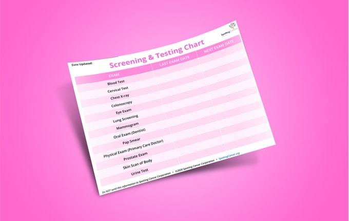
Get the Screening & Testing Scheduling chart
Download the interactive Screening & Testing Scheduling chart to help you keep track of important screening and testing schedules. Download today!
Save a Body Monitoring and Screening & Testing schedule
Regular monitoring and testing is a life-saving habit. Save a Body Monitoring and Screening & Testing schedule to your Google Calendar or iCalendar to stay on track!
You're on Step 5

Step 1:
Signs & Symptoms
To monitor yourself for early cancer detection, you must know the cancer signs and symptoms. A listing of the various signs and symptoms are just a click away.

Step 2:
Body Monitoring
Cancer grows 24/7. Therefore, you must monitor your body to detect any abnormality between regular doctor visits or screenings. The tools and methods are described in this section.

Step 3:
Family History
Knowing and charting your family medical history will help your medical team as they develop a long-term wellness program suited to your unique needs.

Step 4:
Medical Team
Cancer is not self-healing. Therefore, when spotting a cancer sign or symptom, consider it a red flag that should cause you to consult your medical team immediately to determine if it is cancer or another illness.

Step 5:
Screening & Testing
Not all cancer signs and symptoms are visible. You should establish specific times for the various cancer screening and tests with your medical team.


